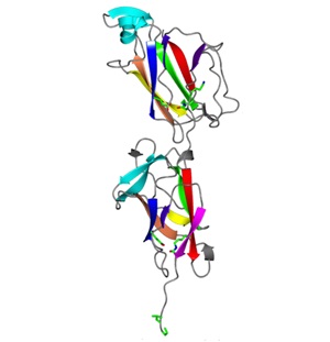
New insights about pilus formation in a probiotic bacterium
Bacterial attachment to the host surfaces is the first and key step in colonization, which may harm or benefit the host based on the natureof host-microbial relationship. Bacteria often assemble and use hair-like organelles known as pili or fimbriae on their cell surface to quickly and effectively mediate attachment. This initial recognition between the bacteria and host through surface molecules determines the tissue tropism and defines host range. Combating bacterial infections by targeting pili or pili-mediated interactions is recognized as a promising approach that may help overcome their ever-increasing repertoires of resistance mechanisms. This requires knowledge of how bacteria assemble pili and use them for adherence. In pathogens, the pili and their components have been studied and also recognized as virulence factors and good vaccine candidates because of their key role in pathogenesis and immunogenic properties.
Interestingly, the beneficial or probiotic bacteria that benefit us also use pili to grab the human gut surface and compete for adhesion with harmful microbes, thus excluding them. Dr. Vengadesan Krishnan’s structural biology research group has begun structural investigations towards understanding how the beneficial bacteria have evolved to utilize pili to adapt the gut environment for survival. As part of their ongoing research, Dr. Krishnan’s group has recently discovered the crystal structure of a building block (backbone pilin) that forms the pilus fiber in the probiotic bacterium Lactobacillus rhamnosus GG. This study presents the first pilin structure from a beneficial bacteria or probiotics and provides new insights about pilus formation. A unique combination of isopeptide bonds and shape complementarity within and between the blocks allow this bacterium to assemble an elongated spring-like pilus fiber. This work contributes to the understanding of how a beneficial probiotic microbe assembles stable pili with the help of isopeptide bonds to withstand environmental shear forces and colonize the gut. The authors further show that the autocatalytic isospeptide bond formation takes place even in the absence of essential catalytic residue.
For more details, https://www.ncbi.nlm.nih.gov/pubmed/27349405

Archives
- January 2024
- December 2023
- October 2023
- September 2023
- July 2023
- November 2022
- September 2022
- June 2022
- April 2022
- March 2022
- January 2022
- December 2021
- August 2021
- July 2021
- May 2021
- April 2021
- February 2021
- January 2021
- December 2020
- October 2020
- September 2020
- August 2020
- July 2020
- June 2020
- April 2020
- February 2020
- January 2020
- December 2019
- November 2019
- October 2019
- September 2019
- May 2019
- April 2019
- January 2019
- December 2018
- July 2018
- June 2018
- May 2018
- April 2018
- February 2018
- January 2018
- November 2017
- September 2017
- June 2017
- February 2017
- January 2017
- April 2016
- January 2016
- September 2015
- July 2015
